A Patient’s Guide to Distal Radius Fractures
What Is A Wrist Fracture?
Welcome back to another one of our ‘intro series’ articles on common hand and upper extremity conditions. As always, if you want the deep dive, you can find that here.
Today we are talking about wrist fractures. Specifically, a distal radius fracture. While this is by far the most common type of wrist fracture, please do confirm with your doctor what type of wrist fracture you have. If you are told distal radius fracture, read on. If you hear words like scaphoid, triquetrum, or hamate, those are separate wrist fracture topics altogether. My ‘intro series’ on scaphoid fractures can be found here.
What Is A Distal Radius Fracture?
A distal radius fracture is the most common type of ‘wrist fracture.’ While the radius is actually a forearm bone (see Figure 1), it commonly breaks near the junction between the forearm and wrist. We call this the ‘distal’ end of the bone (opposite of ‘proximal,’ which is near your elbow) — this is where the term distal radius fracture comes from.
Oh, and before we get too far. Contrary to popular belief, there is no difference between the terms ‘fracture’ or ‘break’. Zero. None. Truly. So I will use the terms interchangeably.
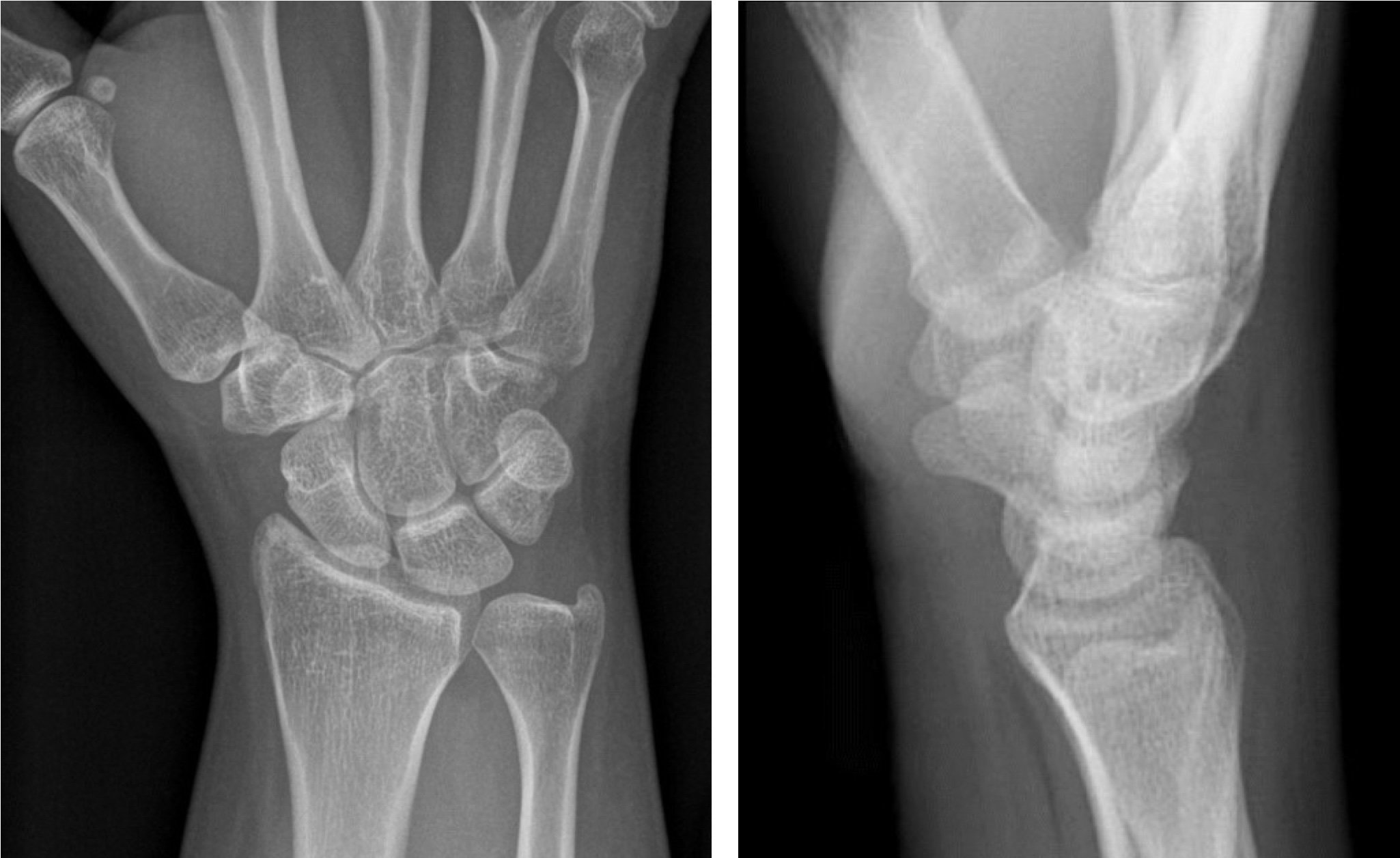
Below you will see x-rays of a normal wrist in Figure 1, followed by x-rays of a distal radius fracture in Figure 2. Can you spot the break?
Figure 1 - Wrist x-rays of a normal (not broken) distal radius
Figure 2 - Wrist x-rays showing a distal radius fracture
Did you see the fracture?
If you are having trouble, look at Figure 3 below where I have outlined the fracture in yellow.
Figure 3 - Yellow outline of the two separate radius fracture fragments. Notice on the righthand picture that the yellow fragment on top (more distal) has tilted backward and shortened.
What I want you to keep in mind are the two terms displacement and angulation. Displacement means ‘how far have the two broken bone ends moved away from each other.’ And angulation would be ‘how far angled or out of alignment have they gone.’ There are so many details to get caught up with when analyzing wrist x-rays, but 99% of your understanding will hinge (see what I did there) on these two concepts.
Let’s spend one more moment thinking about that. Why do displacement and angulation matter? Well, if your bones are displaced too much, they may have trouble healing or staying lined up on their own (or in a cast). And if they are angled too far, they may heal but they will heal crooked. Crooked healing leads to various outcomes such as a visible deformity, loss of movement, weakness, or painful arthritis.
Ok, back to the quick learning.
Please reference Figure 4 below. This is a normal wrist x-ray. On the left, notice the angle formed by the red lines, as well as how flat the green line is between the corner of the radius bone and the corner of the ulna bone.
And on the right is the most important image of them all. My patients know I refer to this as the ‘teacup’ image. Notice the yellow ‘U’ or ‘teacup’ that points directly to the ceiling or even a little tipped toward the thumb. This is the north star of wrist alignment. Refer back to Figure 1 to see if you can trace these lines out on your own.
Figure 4 - A normal wrist AP and lateral set of x-rays for comparison to Figure 4.
Do I Need Surgery For My Distal Radius Wrist Fracture?
Now take your new knowledge of what normal wrist x-rays look like and overlay that with Figure 5 below. This is the distal radius fracture. Notice the broken bones have displaced enough to dramatically shrink the angle between the red lines and move the corner of the distal radius far below the corner of the ulna. And what happened to our teacup? That thing is now pointed 45 degrees backwards! Angulation, angulation, angulation…
Figure 5 - A distal radius fracture demonstrating shortening (compare green arrow and the angle between the two red lines to Figure 5) on the AP x-ray. The lateral x-ray demonstrated the backward or ‘dorsal’ tilt of the fractured distal radius fragment.
And as I mentioned above, displacement and angulation lead to poor bone healing, crooked bone healing, weakness, stiffness, arthritis…all kinds of bad things. Suffice it to say that if your x-rays look like Figure 6, you are going to want to consider surgery to return your displacement and angulation closer to normal.
What Is The Surgery For A Distal Radius Fracture?
I have more information in my in-depth article here, but the basic answer is with a plate and screws. This is called ‘open reduction internal fixation’ or ORIF in the orthopedic world.
Translation: Open the skin (surgery). Reduce the fracture (put it back in alignment). And place something Internally (plate and screws) to Fix it in place.
Figure 6 - AP and lateral x-ray of a distal radius fracture after a volar-locked plate has been used to surgically fix the fracture.
Figure 7 - Compare the yellow, green, and red annotations to Figures 4 and 5. The angulation (yellow arrow) and height (green arrow & angle between the red lines) of the distal radius fracture have been restored to the normal anatomic range.
Figure 6 is an x-ray of a distal radius fracture that has now been fixed with a plate and screws. And Figure 7 draws the lines you will remember from our earlier x-rays.
Notice how with surgery, I have increased the angle between the red lines back to normal, restored the relationship between the corner of the radius and the corner of the ulna (green line), and put that darn teacup back up on top where it belongs (yellow).
So there you have it. Now you know a little more about wrist fractures, why we fix them, and how we fix them with surgery. As I’ve implied before, you can imagine the endless nuance around this topic, but that is for another day…


